
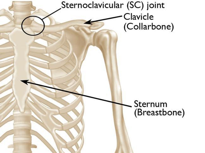
The sternoclavicular joint is located between the collarbone (clavicle) and the breastbone (sternum), and it is the only joint that connects the arm to the trunk.
Add us as a preferred source on GoogleSee our new articles higher in your searchesAddIt allows the shoulder girdle to move in coordination with the body.
Despite its small size, the joint is critically important for upper extremity functions.
Immediately behind the sternoclavicular joint lie the trachea, esophagus, and major blood vessels leading to the head and neck.
For this reason, in cases where the sternoclavicular joint dislocates backward (posterior dislocation), not only are shoulder movements restricted, but life-threatening risks such as breathing, swallowing, or circulation problems may also occur.
Acute sternoclavicular joint dislocations are reduced in a hospital setting.
If permanent instability develops in the joint, surgery may be required.
In this case, the surgery usually involves a reconstruction procedure in which the ligaments are rebuilt.
Similar to an anterior cruciate ligament (ACL) reconstruction in the knee, one of the patient’s hamstring tendons (most commonly the gracilis) is harvested and used as a graft to restore stability of the joint.
What Is the Sternoclavicular Joint?
The sternoclavicular joint (SCJ) forms the connection between the collarbone (clavicle) and the breastbone (sternum).
As the only joint that links the shoulder girdle to the trunk, it is indispensable for weight-bearing, arm movements, and synchronized motion with the torso.
The capsule and ligaments surrounding the joint provide both mobility and stability.
However, when exposed to trauma, these structures may be damaged, leading to dislocation or instability.
Although dislocations are rare, posterior dislocations in particular can pose life-threatening risks.
What Are Sternoclavicular Joint Injuries?
Sternoclavicular joint injuries usually occur as a result of trauma.
These injuries may present as a dislocation or a subluxation (partial dislocation).
The direction of the dislocation is important, because whether the clavicle is displaced forward (anterior) or backward (posterior) changes both the symptoms and the risks.
- Anterior dislocation (forward displacement of the clavicle) is more common. It usually presents with obvious swelling and deformity.
- Posterior dislocation is less common but much more serious. Because just behind the sternoclavicular joint lie major blood vessels leading to the heart and lungs, as well as the esophagus and trachea, posterior dislocations can compress these structures and cause breathing and circulatory problems.
Traumatic dislocations generally result from high-energy injuries, such as traffic accidents or contact sports (e.g., football, rugby).
In patients younger than 20–25 years old, sternoclavicular dislocation can be confused with a separation of the clavicle from the growth plate (physis), since in this age group the physis has not yet fully closed and may appear similar.
In some individuals, spontaneous subluxations can occur without trauma when the arm is raised.
These episodes are often seen in younger patients and are associated with ligamentous laxity (looseness of the ligaments).
The subluxation typically resolves spontaneously when the arm is lowered.
In such cases, surgery is usually unnecessary; reassurance of the patient and treatment aimed at local symptoms are sufficient.
Symptoms of Sternoclavicular Joint Injuries
The symptoms of sternoclavicular joint injuries vary depending on the direction of the dislocation and whether chronic instability has developed.
Patients typically present with pain around the clavicle, restricted shoulder movements, and a feeling of abnormal mobility of the bone.
Symptoms of Sternoclavicular Joint Dislocation (Acute Dislocation)
Sternoclavicular joint dislocations that occur after trauma present with sudden symptoms. The most common findings are:
- Anterior dislocation (forward displacement): A prominent bony lump is visible under the skin. Tenderness and swelling occur with palpation. Shoulder movements are painful.
- Posterior dislocation (backward displacement): A visible deformity may not always be present. However, difficulty swallowing, shortness of breath, chest pressure, or hoarseness may develop. Because posterior dislocations compress the vital vessels and organs behind the sternum, they require emergency intervention.
Symptoms of Chronic Sternoclavicular Joint Instability
In some patients, the joint loses stability after the initial trauma, leading to chronic sternoclavicular instability. This can significantly affect quality of life, especially in active individuals.
Common symptoms of chronic instability include:
- Forward–backward movement of the clavicle over the sternum during shoulder motion
- A shifting sensation or clicking sound in the anterior chest wall
- Increased discomfort and loss of confidence when lifting the arm
- Recurrent pain or pressure with daily movements
- Cosmetic deformity or asymmetry at shoulder level
Sternoclavicular joint instability is often overlooked at first glance.
However, in patients with long-term symptoms and a feeling of abnormal motion at the clavicle during activity, the correct diagnosis must be made, and surgical planning considered if necessary.
How Are Sternoclavicular Joint Injuries Diagnosed?
Accurate diagnosis of sternoclavicular joint dislocations and instabilities is essential for effective treatment planning.
Posterior dislocations and chronic instability, in particular, may not be obvious on physical examination alone. For this reason, both clinical assessment and imaging methods must be used.
Physical Examination
During physical examination, the physician may detect:
- Swelling, tenderness, or deformity at the level of the clavicle
- Pain or abnormal motion with arm movements
- In suspected posterior dislocation: swallowing difficulty or breathing problems
However, posterior dislocations may be missed on physical exam. Therefore, imaging is critical.
Imaging Methods
X-ray
Standard anteroposterior radiographs are usually requested first.
However, because the sternoclavicular joint overlaps with other bony structures, the exact direction of dislocation is not always clear on plain X-rays.
Computed Tomography (CT)
CT is the most valuable imaging method for diagnosing sternoclavicular joint injuries.
It allows three-dimensional evaluation and is essential for determining the direction of the dislocation (anterior vs. posterior).
CT also provides detailed information on joint surface fractures, signs of instability, and relationships with adjacent anatomical structures.
In posterior dislocations, CT is vital for assessing the distance to mediastinal organs and any compression on them. For this reason, CT must always be performed in suspected posterior dislocations.
Magnetic Resonance Imaging (MRI)
MRI provides information about the joint capsule, ligaments, and surrounding soft tissues.
However, CT remains the first-line diagnostic tool.
MRI is more useful for treatment planning or in cases of suspected chronic instability.
Treatment Methods for Sternoclavicular Joint Injuries
The treatment plan for sternoclavicular joint injuries depends on the direction of the dislocation, associated structural damage, and whether permanent instability has developed.
Acute Sternoclavicular Joint Dislocation Is an Emergency!
Especially posterior dislocations carry the risk of compressing vital organs and therefore require urgent intervention.
In such cases, under hospital monitoring, the joint is reduced (closed or open reduction) by experienced specialists.
Anterior dislocations are usually less risky and do not pose an immediate life-threatening danger.
However, these dislocations can still cause severe damage to the joint capsule and may lead to instability over time.
Treatment of Chronic Sternoclavicular Joint Instability
If the joint capsule and ligaments do not heal properly after the initial trauma, chronic sternoclavicular instability may develop.
This condition can cause significant functional loss and reduced quality of life, especially in active individuals.
A constantly unstable clavicle creates both physical and psychological discomfort.
If the complaints due to instability are mild, physical therapy and strengthening exercises for the shoulder girdle may be tried.
However, in many patients, these methods are insufficient.
Who Needs Surgery for Chronic Sternoclavicular Joint Instability?
Surgery is required in chronic sternoclavicular instability, particularly in the following situations:
- Recurrent dislocations or marked joint laxity
- Pain and instability interfering with daily activities
- Functional limitation in active patients
- Posterior chronic instability causing life-threatening symptoms such as swallowing difficulty or breathing problems
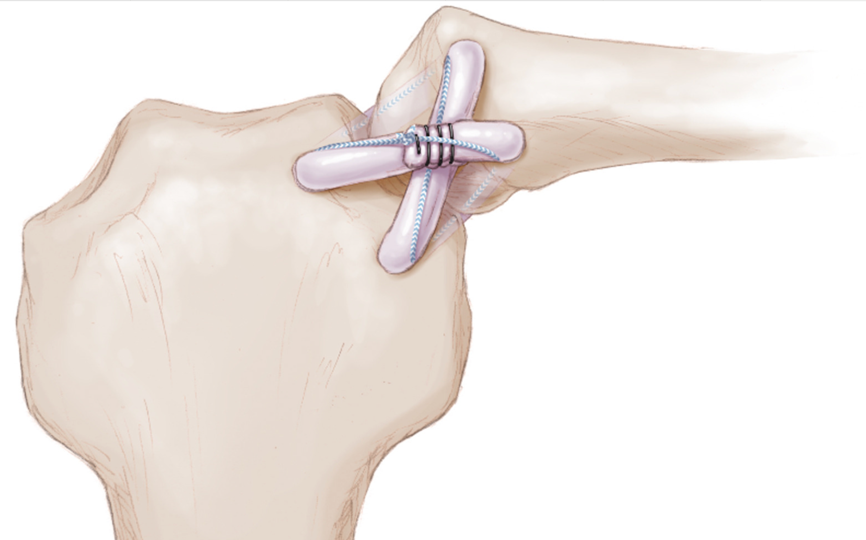
In reconstruction surgery, a tendon graft is used to replace the damaged ligaments.
Most commonly, the gracilis tendon harvested from the patient’s hamstring is preferred.
The graft is passed through tunnels created between the clavicle and the sternum, reconstructing the ligaments and restoring stability of the joint.
The success rate after surgery is high, and when performed correctly, most patients can safely return to daily and sports activities.
Sternoclavicular Joint Reconstruction Surgery
The permanent solution for chronic sternoclavicular joint instability is surgical reconstruction.
The aim of this procedure is to restore joint stability by replacing the loosened or torn ligaments with a graft.
The gracilis tendon from the patient’s hamstring group is usually used as the graft.
This tendon is placed between the clavicle and sternum with special techniques.
Passed through tunnels drilled into the bones, the graft is fixed in a way that replicates the function of the original ligaments.
Thus, the sternoclavicular joint is stabilized and balanced again.
In posterior instabilities, the positioning of the graft is carefully planned in relation to vital structures.
If necessary, reconstruction is performed with an open surgical technique.
- Joint stability is restored,
- The abnormal feeling of looseness disappears,
- Chronic pain, swallowing, or breathing difficulties are significantly improved,
- Patients can safely return to daily life and sports.
When performed with the correct technique by an experienced surgeon, reconstruction surgery has a high success rate.
Postoperative Process and Rehabilitation
The recovery process after sternoclavicular joint reconstruction surgery progresses step by step and in a controlled manner.
Rehabilitation is crucial to protect the success of the surgery and ensure proper healing of the graft.
First 4 Weeks: Immobilization Phase
The arm is kept immobilized with a shoulder sling for at least 4 weeks to restrict movement.
Only passive and safe exercises, such as pendulum movements, are allowed.
The goal is to allow soft tissues to heal without excessive stress on the joint.
After 4 Weeks: Range of Motion and Strengthening
From the fourth week, patients begin active-assisted exercises.
Initially, shoulder abduction (lifting the arm sideways) is limited to 90 degrees.
Depending on the clinical progress, range of motion is gradually increased.
Weight-bearing and strengthening exercises are introduced step by step under the supervision of the surgeon and physiotherapist.
After 12 Weeks: Daily Activities and Return to Sports
Return to high-level physical activity or sports is usually considered no earlier than 12 weeks postoperatively.
This timing is individualized according to the patient’s joint stability, pain-free mobility, and muscle strength.
Frequently Asked Questions About Sternoclavicular Joint Dislocation
What Is the Sternoclavicular Joint and What Does It Do?
The sternoclavicular joint is located between the collarbone (clavicle) and the breastbone (sternum). It is the only joint that connects the arm to the trunk and allows the shoulder girdle to move in coordination with the body.
Is Sternoclavicular Joint Dislocation Dangerous?
Yes, especially posterior dislocations are dangerous. They may compress the trachea, esophagus, and major blood vessels, leading to breathing or circulatory problems.
What Is the Difference Between Posterior and Anterior Dislocations?
In an anterior dislocation, the clavicle shifts forward and usually does not pose a life-threatening risk. In a posterior dislocation, the clavicle moves toward the chest cavity and may press on vital structures, which can be life-threatening.
How Can Chronic Sternoclavicular Joint Instability Be Recognized?
Typical signs include forward–backward movement of the clavicle during shoulder motion, clicking or popping sensations, recurrent pain, and a feeling of instability around the shoulder.
What Happens If Chronic Instability Is Not Treated?
Pain may worsen over time, movement restriction can develop, and in posterior instability, symptoms such as swallowing or breathing difficulties may occur. Long-term, quality of life is significantly reduced.
When Is Surgery Necessary?
Surgery is required in cases of recurrent dislocations or marked laxity, pain or instability interfering with daily life, and posterior instability associated with life-threatening symptoms.
How Is Sternoclavicular Joint Surgery Performed?
A reconstruction procedure is performed, in which a graft (usually the gracilis tendon from the patient’s hamstring) is placed between the clavicle and sternum to restore stability.
How Long Must the Arm Remain in a Sling After Surgery?
The arm is generally immobilized in a sling for about 4 weeks. Only passive exercises such as pendulum movements are allowed during this period.
When Can Patients Return to Sports or Heavy Work?
Return to high-demand physical activity is usually considered 12 weeks after surgery, depending on individual recovery.
What Is the Success Rate of This Surgery?
When performed with the correct technique by experienced surgeons, the success rate is high. Most patients return to daily life with a pain-free and stable shoulder.
Conclusion
Sternoclavicular joint injuries occur at the joint connecting the clavicle to the sternum.
Posterior dislocations after trauma are emergencies because they can compress the trachea and major vessels, creating life-threatening risks.
More common anterior dislocations usually cause chronic instability and recurrent pain rather than immediate danger.
When the initial injury fails to heal and persistent instability develops, the definitive solution is surgical reconstruction.
In this procedure, a graft taken from the patient’s hamstring tendon restores stability, allowing the patient to return to daily life safely and without pain.
Expert Recommendations
In suspected sternoclavicular dislocation, especially after high-energy trauma, accurate diagnosis is critical.
Posterior dislocations must not be overlooked. CT scanning is the most reliable imaging method.
Symptoms such as shortness of breath, difficulty swallowing, or hoarseness require immediate emergency care.
Not all dislocations require surgery. Surgical treatment is mainly considered in cases of persistent painful instability affecting daily life.
Successful outcomes depend not only on surgical technique but also on strict adherence to the postoperative rehabilitation program.
For a personalized evaluation and to determine the most appropriate treatment, you can contact Utku Erdem Özer directly.
For more details, please use the contact channels or book an appointment.