
Piriformis syndrome is a muscle-related disorder that occurs when the piriformis muscle presses on the sciatic nerve running just beneath it, causing pain, numbness, and tingling in the hip and leg, and it is frequently confused with a lumbar disc herniation. It is seen especially in people who sit for long periods, desk workers, and athletes who load one side of the body.
Add us as a preferred source on GoogleSee our new articles higher in your searchesAddIn medicine it is sometimes also called extradiscogenic sciatica — that is, sciatic pain that does not come from the disc. Here, the term “not from the disc” conveys this: the source of sciatica-type pain is not always the disc in the lower back; in other words, the problem does not necessarily have to be a lumbar disc herniation or lumbar degeneration. Sometimes the source of the pain is much lower down, in a muscle deep in the buttock. If your lumbar MRI comes back perfectly clean yet you have persistent pain radiating from the hip down the leg, piriformis syndrome is a strong diagnostic candidate.
During the examination, my patients describe this picture with a very typical sentence: “They told me my lumbar MRI is clear, but I still can’t sit; there’s a pain shooting from my hip down my leg.” In this article, I explain—through the eyes of an orthopedic specialist and endurance athlete—the symptoms of piriformis syndrome, how I distinguish it from a lumbar disc herniation, how I make the diagnosis, and all the steps from non-surgical treatment to surgery.
📋 Quick Summary
Piriformis syndrome occurs when the piriformis muscle deep in the buttock presses on the sciatic nerve. Its most characteristic symptom is the inability to sit for long periods: after sitting for 15-20 minutes, especially on a hard surface, the pain becomes unbearable and the patient constantly changes position.
It is very often confused with a lumbar disc herniation. The key to distinguishing them: if the lumbar MRI is clear and pain radiating into the leg persists, the piriformis is considered—because piriformis syndrome is a diagnosis of exclusion and usually does not show up on MRI.
Treatment always begins with non-surgical methods: activity modification, stretching exercises, and, when needed, ultrasound-guided (US) injection. Early cases mostly resolve within 4-8 weeks. Surgery only comes onto the agenda in resistant cases lasting longer than 3 months that do not respond to treatment, and it leaves no loss of function.
What Does Piriformis Mean?
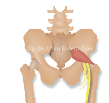
The piriformis is a muscle located behind the hip joint, deep in the gluteal (buttock) region. Its name comes from the Latin pirum, meaning “pear,” and forma, meaning “shape”; in other words, it means the pear-shaped muscle.
The region where this muscle sits is called the deep gluteal space. The job of the piriformis is to rotate the leg and hip outward (external rotation)—and it is not the only muscle that does this; there are 6 other muscles that share the same task. Keep this detail in mind; when surgery comes onto the agenda, we will return to it to explain why there is no loss of function.
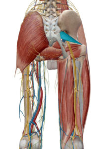
The piriformis muscle originates from inside the pelvis (the front surface of the sacrum) and attaches to the upper prominence of the hip bone (the greater trochanter). During this course, it crosses diagonally over the sciatic nerve. The whole story lies in this proximity: when the muscle enlarges and tightens, it compresses the thickest nerve lying right beneath it.
Is your lumbar MRI clear but your pain won’t go away?
Correctly identifying the source of pain radiating from your hip down your leg is the first step to avoiding unnecessary treatments. Get in touch for an evaluation.
What Is Piriformis Syndrome?
Piriformis syndrome is the condition that occurs when the piriformis muscle enlarges (hypertrophies) or goes into spasm—that is, tightens and locks—and presses on the sciatic nerve just beneath it. Especially when the muscle works more than normal and incorrectly because of sports activities or repetitive movements at work, it tightens; pain begins in the hip and thigh region, and in some patients numbness extends down to the heel. The condition usually becomes prominent after prolonged sitting, incorrect training, or unbalanced muscle use.
So how common is it? Piriformis syndrome is responsible for a relatively small but important portion of sciatica-type pain radiating into the leg; research shows that in about 5-6% of patients presenting with low back and leg pain, the source of the pain may be the piriformis. It most often appears between the ages of 40 and 50 and more in women than in men. This difference in frequency makes recognizing the condition even more important: because when it is not kept in mind enough, it can easily be brushed off with a “lumbar disc herniation” label.
In some patients, the condition begins with a trauma such as falling on the buttock; a sudden strain triggers an injury in the muscle and then a process of pressure on the nerve. As an endurance athlete, another scenario I frequently see is one-sided loading—one-sided rowing is the best example of this.
In some people, the problem is a congenital anatomical variation. In about one-fifth of the population, the sciatic nerve passes not beneath this muscle but through it, or splits in two and wraps around both above and below the muscle. In these individuals, because every contraction of the muscle directly compresses the nerve, the predisposition to this condition is greater and the symptoms can be more stubborn.
Here I would like to add an important point: the piriformis is not the only structure that compresses the sciatic nerve deep in the buttock. The nerve can also come under pressure from other muscles, bands of connective tissue, or blood vessels in the area we call the deep gluteal space deep in the buttock. In current medicine, this entire picture is called deep gluteal syndrome; piriformis syndrome is its most common and best-known subtype. So instead of saying “the problem is always in this muscle,” looking at it as “a nerve is being compressed somewhere deep in the buttock” allows us to catch causes that are missed in diagnosis.
What Are the Causes of Piriformis Syndrome?
Piriformis syndrome is not due to a single cause; most of the time several factors come together. The most common causes I encounter are:
- Sitting for long periods. The biggest trigger of our time. In office workers and drivers, the muscle stays under pressure for hours and tightens.
- “Wallet sciatica.” A thick wallet carried in the back pocket presses directly on this muscle and the sciatic nerve when you sit. This condition, popularly known by this name, is surprisingly common—and its solution is sometimes simply moving the wallet to the front pocket.
- Repetitive, one-sided, or excessively straining sports. Incorrect or excessive loading of the muscle in activities such as running, cycling, or one-sided rowing.
- Direct trauma. Muscle injury after falling on the hip, followed by spasm.
- Anatomical difference. Congenital variations in which the sciatic nerve passes through or around the muscle.
- Biomechanical imbalance. In conditions such as scoliosis or a leg-length discrepancy, the piriformis on one side works excessively and enlarges (hypertrophy).
- Starting sports without warming up and tight, snug clothing—habits that create microtrauma in the muscle.
What Are the Symptoms of Piriformis Syndrome?
The symptoms of piriformis syndrome follow the path of the compressed sciatic nerve. The earliest finding is usually numbness or tingling in the buttock; pain is usually added later, gradually. Typical symptoms are:
- Pain deep in the buttock — over time it can spread to the back of the thigh, the back of the knee, and in some patients down to the heel.
- Inability to sit for long periods — most patients say they cannot sit for more than 15-20 minutes, especially on a hard surface. This is the most characteristic signature of the condition.
- Pain and numbness that increase when moving the hip and leg (especially when rotating inward).
- A sensation of numbness throughout the leg on waking in the morning.
- Pain triggered by sitting, running, or climbing stairs or slopes.
- Discomfort that becomes prominent when crossing the legs.
- Spasm and stiffness that the doctor feels by hand in the muscle during the examination.
During the examination, my patients very often sum it up like this: “After sitting on a hard chair for 10 minutes, an unbearable pain starts in my buttock; I constantly change position; I can’t sit on the painful side.” When I hear this sentence, one of the first diagnoses that comes to mind is the piriformis.
I would also like to add: piriformis syndrome does not always behave “by the textbook.” In some patients, the pain is not limited to the hip and leg; less well-known symptoms can also occur, such as tingling in the foot, a sensation of pressure/fullness in the groin or pelvic region, and even discomfort that increases when sitting on the toilet or on a bicycle saddle. These atypical symptoms make it easier to confuse the condition with other disorders. For example, if there is pain radiating into the groin, our article on groin pain may also help in distinguishing its source.
If you have severe hip pain and are wondering about its source, you can also take a look at our article on femoroacetabular impingement syndrome; it is a condition that produces pain in a similar region but through a different mechanism.
🚩 When should you see a doctor without losing time?
Piriformis syndrome usually eases with non-surgical methods; however, the following symptoms may be the herald of a different and more serious nerve problem. If you have any of these, you need to be evaluated without delay:
- Loss of control of urination or bowel movements (incontinence or inability to go)
- Sudden dropping of the foot, inability to lift the foot off the ground (foot drop)
- Rapidly progressing, increasing weakness or loss of sensation in the leg
- Numbness on the inner side of both legs / in the groin-perineal region (“saddle” numbness)
- Frequent falls due to pain or numbness
These symptoms suggest an emergency picture originating from the spine or nerve root rather than piriformis syndrome; in such a situation, no time should be lost.
What Is the Difference Between Piriformis Tightness and Piriformis Syndrome?
Piriformis tightness and piriformis syndrome are most of the time thought to be the same; yet the difference between them is the most important distinction that determines the course of treatment. The single decisive question is this: is the pain accompanied by sciatic nerve findings or not?
- Piriformis tightness: Pain is felt only locally, deep in the buttock. There is no radiation into the leg, numbness, or tingling. That is, the nerve is not yet involved; the muscle has tightened but is not pressing on the sciatic nerve.
- Piriformis syndrome: The muscle is now pressing on the sciatic nerve. Pain radiates from the hip down the leg; nerve findings such as numbness and tingling are added to the picture. This is a stage beyond tightness.
| Feature | Piriformis Tightness | Piriformis Syndrome |
|---|---|---|
| Location of pain | Only deep in the buttock, local | Radiates from the hip down the leg |
| Nerve findings (numbness, tingling) | Absent | Present |
| Pressure on the sciatic nerve | Not yet | Present |
| Stage | Early phase — herald of the syndrome | Advanced phase |
⏳ Why is early intervention important?
If piriformis tightness is not treated in time, it eventually presses on the sciatic nerve and turns into piriformis syndrome. That is, a condition that was initially only a local muscle pain evolves, as it progresses, into a nerve problem that radiates into the leg and causes numbness. That is why the most valuable window of opportunity is the tightness phase, before neuropathic sciatic pain (that is, nerve-related pain that radiates into the leg) begins.
In this early phase, my ideal treatment approach is this: to relieve the tightness of the muscle with regular stretching exercises and, when needed, to support this with a series of ultrasound-guided injections—usually planned as 3 doses in total, one month apart. The goal is to eliminate the tightness before the muscle presses on the nerve and to prevent the condition from progressing to the syndrome. Which injection is appropriate and the dosing interval are determined individually according to the examination findings.
In short: if you have a local, non-radiating tightness and pain in your buttock, evaluating it early instead of postponing it as “it will pass” can head off a much more difficult process later on.
Is Piriformis Syndrome Confused With a Lumbar Disc Herniation? How Can I Tell the Difference?
Yes, and very often. There are points I pay attention to when distinguishing the two—but first I want to correct a common misconception:
- Don’t rely too much on where the pain starts. It is classically said that “in a lumbar disc herniation the pain starts from the lower back, and in the piriformis it starts from the hip”; but this distinction is not reliable in practice. Because in a lumbar disc herniation the low back pain usually subsides within the first 2 weeks and what remains is the sciatic pain radiating into the leg—so the picture increasingly begins to look like “hip/leg pain.” Moreover, in a lumbar disc herniation, too, the center of the pain can be deep in the buttock. That is why where the pain starts is not decisive on its own.
- Not being able to sit is the most valuable clue. The piriformis patient says “I can’t sit for more than 15-20 minutes; the pain becomes unbearable on a hard surface; I constantly change position.” A marked increase in pain on a hard chair is the most characteristic signature of this condition.
- Clear neurological findings guide the way. Clear neurological findings such as marked loss of sensation, weakness in the toes, and foot drop, along with pain descending below the knee and even to the foot, suggest more a true nerve-root compression—that is, a lumbar disc herniation. Such marked signs of nerve damage are usually not prominent in the piriformis.
So how do we actually make the distinction? The most practical route is imaging—but with an unexpected logic. Keep this in mind: piriformis syndrome is a diagnosis of exclusion. With MRI, we usually cannot directly show the muscle itself; that is, imaging alone does not “prove” the diagnosis. But here is the useful side: if the lumbar MRI comes back clean and the patient still has sciatica-type pain radiating into the leg, this tells us there is no lumbar disc herniation. Once we have ruled out the herniation, the remaining picture strongly suggests this syndrome. In short, MRI shows us what is not present; we complete the rest of the picture with the examination.
How Is Piriformis Syndrome Diagnosed?
Diagnosing piriformis syndrome is not easy; it requires experience. The fact that its symptoms overlap almost exactly with those of a lumbar disc herniation is what makes the diagnosis hardest. In a patient with suspected piriformis, the first thing I do is examine the lower back region and, with MR imaging, show that there is no lumbar disc herniation.
There is no specific blood test or imaging study that directly identifies piriformis syndrome. Since there is no single test that establishes the diagnosis on its own, the diagnosis proceeds with a stepwise logic:
- Causes that produce a similar picture (lumbar disc herniation, spinal canal stenosis, sacroiliac joint problem) are ruled out.
- Special examination tests targeting this muscle are applied; reproduction of the pain supports the diagnosis.
- When needed, a US-guided diagnostic injection is performed; a marked reduction in pain both confirms the diagnosis and initiates treatment.
All of these special tests we use in the examination share a common logic: to temporarily increase pressure on the nerve by stretching or contracting this muscle and to reproduce the familiar pain. Let me describe the ones I use frequently in patient-friendly terms:
- FAIR test (one of the tests I use most often) — While you lie on your side, we flex your painful leg at the hip and knee and rotate it inward. Because this movement stretches the piriformis, if the nerve is being compressed, your familiar pain or a sensation radiating into the leg appears.
- Freiberg test — With your leg extended straight, we rotate your hip inward. If the piriformis is stretched and pain is felt deep in the buttock, the test is meaningful. (It does not come out positive in every patient on its own; it produces pain in about two-thirds of cases.)
- Pace test — While seated, you try to spread your legs outward and we resist this with our hand. Feeling pain or weakness while pushing against this resistance supports the test.
- Beatty test — While you lie on your healthy side, we ask you to raise and hold the upper, painful leg a few centimeters up with the knee bent. Pain arising deep in the buttock points to the piriformis.
A single positive test does not confirm the diagnosis; what really matters is that several tests together reproduce the pain. That is why I evaluate these tests together with your history and other examination findings—just like putting together the pieces of a puzzle.
One detail is important: in only 10-15% of patients with a confirmed piriformis diagnosis is the muscle seen enlarged compared to the opposite side on pelvic MRI. That is, a clean MRI does not rule out the piriformis; this is precisely why it is a “diagnosis of exclusion.”
Piriformis Syndrome MRI Image
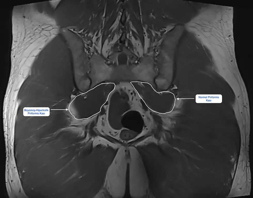
In the pelvic MRI image below, the muscle on the right side is seen to be markedly enlarged (hypertrophic) compared to the opposite side. This is a relatively “lucky” image that we can capture in only a small portion of diagnosed patients—because in most patients the MRI does not show the muscle directly. The patient in this image underwent surgery after a lengthy treatment process and was freed of the complaints.
So what happens when a standard MRI is inadequate? This is where a special imaging technique called MR neurography can come into play. While a standard MRI mostly shows bone and general soft tissue, MR neurography is tuned to image the nerve itself directly; it can reveal signs of thinning, thickening, or compression in the sciatic nerve. It is not needed for everyone and is not available at every center; however, it can be a valuable guide in resistant cases where the diagnosis is unclear and that do not respond to non-surgical treatment. Even so, I would like to underline: even the most advanced imaging does not replace the examination—it complements it.
How Is Piriformis Syndrome Treated?
There is no single method in treating this syndrome, and I always start treatment from the least demanding step. What will work in which patient is largely determined by how long the condition has been going on. The steps are as follows:
1. Non-surgical treatment — always the first step
- Activity modification and relative rest: Taking a break from prolonged sitting, one-sided loading, and strenuous sports that trigger the pain. If there is “wallet sciatica,” even changes as simple as moving the wallet to the front pocket make a difference.
- Stretching and strengthening exercises: A program guided by a physiotherapist that stretches the muscle and strengthens the weak hip muscles. It is the cornerstone of treatment.
- Manual therapy: Deep-tissue and trigger-point techniques applied by an experienced physiotherapist.
- Medication support: Painkillers and muscle relaxants when needed—to provide enough relief to be able to exercise.
2. Interventional treatment — US-guided injections
If non-surgical methods do not provide sufficient relief, the next step is injections. The most critical point here is this: piriformis injections must be placed precisely into the muscle under ultrasound (US) guidance, not blindly. Because the muscle is deep in the buttock, it is hard to reach the correct point without imaging. I have compared the options in a separate table in the section below.
The first injection I prefer for the piriformis is a US-guided cortisone injection: cortisone given together with a local anesthetic provides rapid relief by suppressing the swelling and inflammation in the muscle and the inflammation in the irritated nerve. This window of relief is very valuable; because once the pain eases, the patient can perform stretching and strengthening exercises efficiently. In cases that do not get sufficient benefit from cortisone and that have resistant and recurrent spasm, Botox—the injection with the strongest evidence in current research—comes onto the agenda. In selected cases that do not respond to established treatments, PRP, which targets tissue repair, may be an option. I have compared these three options, together with their levels of evidence, in a separate table below.
3. Surgery — only the very last step
The first phase of treatment is always non-surgical methods. However, in resistant cases lasting longer than 3 months that do not respond to physical therapy and seriously reduce quality of life, surgery comes onto the agenda. I have explained the decision for surgery and its details in a separate section below.
Expectations by duration: While recently started cases get rapid benefit from stretching, manual therapy, rest, and injections, in chronic cases the benefit obtained from these applications is limited or temporary. That is why, when starting treatment, how long the condition has been going on is one of the most decisive questions.
Piriformis Injections: The Difference Between Cortisone, Botox, and PRP
When we say “injection,” don’t picture a single needle; there are different options with different purposes, durations of effect, and levels of evidence behind them. I have summarized which is suitable for whom in the table. The common rule does not change: all of them are done under US guidance, precisely into the muscle—because since the muscle is deep in the buttock, it is hard to reach the correct point without imaging.
| Method | What does it do? | Duration of effect | Who is it suitable for? |
|---|---|---|---|
| Cortisone injection | A mixture of local anesthetic + cortisone; suppresses the swelling and inflammation in the muscle and the irritation in the nerve | Starts quickly, lasts relatively briefly (weeks) | The patient whose pain is prominent and who needs rapid relief in order to be able to start exercising |
| Botox (botulinum toxin) | Temporarily relaxes the muscle; targets the muscle-based mechanism of the problem by stopping the excessive contraction and the pressure on the nerve | Longer (about 3-6 months) | The patient with resistant, recurrent spasm who does not get sufficient/lasting benefit from cortisone |
| PRP (platelet-rich plasma) | Prepared from the person’s own blood; contains growth factors that support tissue repair beyond just suppressing inflammation | Gradual; repair-oriented | A selected resistant case that does not respond to established treatments (exercise, cortisone, Botox) and in whom healing at the tissue level is targeted |
I would like to expand a little on these three options in light of current research, because the difference between them is not just “duration of effect” but the level of evidence behind them:
- Cortisone — It is the most frequently used option in daily practice; its effect starts quickly but is relatively short-lived. Because it rapidly reduces pain and gets the patient to a state where they can exercise, it is my first choice especially in cases where the pain is prominent.
- Botox — Among injection treatments in piriformis syndrome, it has the strongest level of evidence. Controlled studies and the large-scale analyses that combine them show that Botox can give superior results in reducing pain compared to a cortisone + local anesthetic mixture. Its logic is also clear: Botox directly resolves the excessive muscle contraction at the source of the problem, thereby relieving the pressure on the nerve. That is why it is a strong option in resistant, recurrent spasms.
- PRP — It is a method obtained from the person’s own blood that supports tissue repair. The evidence for PRP in piriformis syndrome is still new and limited; however, the first controlled study conducted showed that US-guided PRP provided significant improvement in pain and function. For this reason, I consider PRP not as a routine first step, but as an option for selected resistant cases that do not respond to established treatments (exercise, cortisone, Botox) and in which I target healing at the tissue level. The goal is not just to suppress the pain but to support the repair of the tissue.
As you can see, there is no single “best injection”; the right choice varies according to the patient’s clinical picture, how long the complaint has been going on, and the response to previous treatments. Which injection is appropriate is determined individually, together with the examination findings. For broader information about injection options, you can review our injection treatments page.
One purpose of injections is also to move the patient into a pain-free window for physical therapy: when the muscle is relaxed, stretching and strengthening are much more efficient. That is why I view an injection not as a “magic” on its own, but as a tool that makes the exercise program possible. I would also like to remind you: in chronic cases, the likelihood of injections producing results on their own is low.
Piriformis Syndrome Exercises and What to Avoid
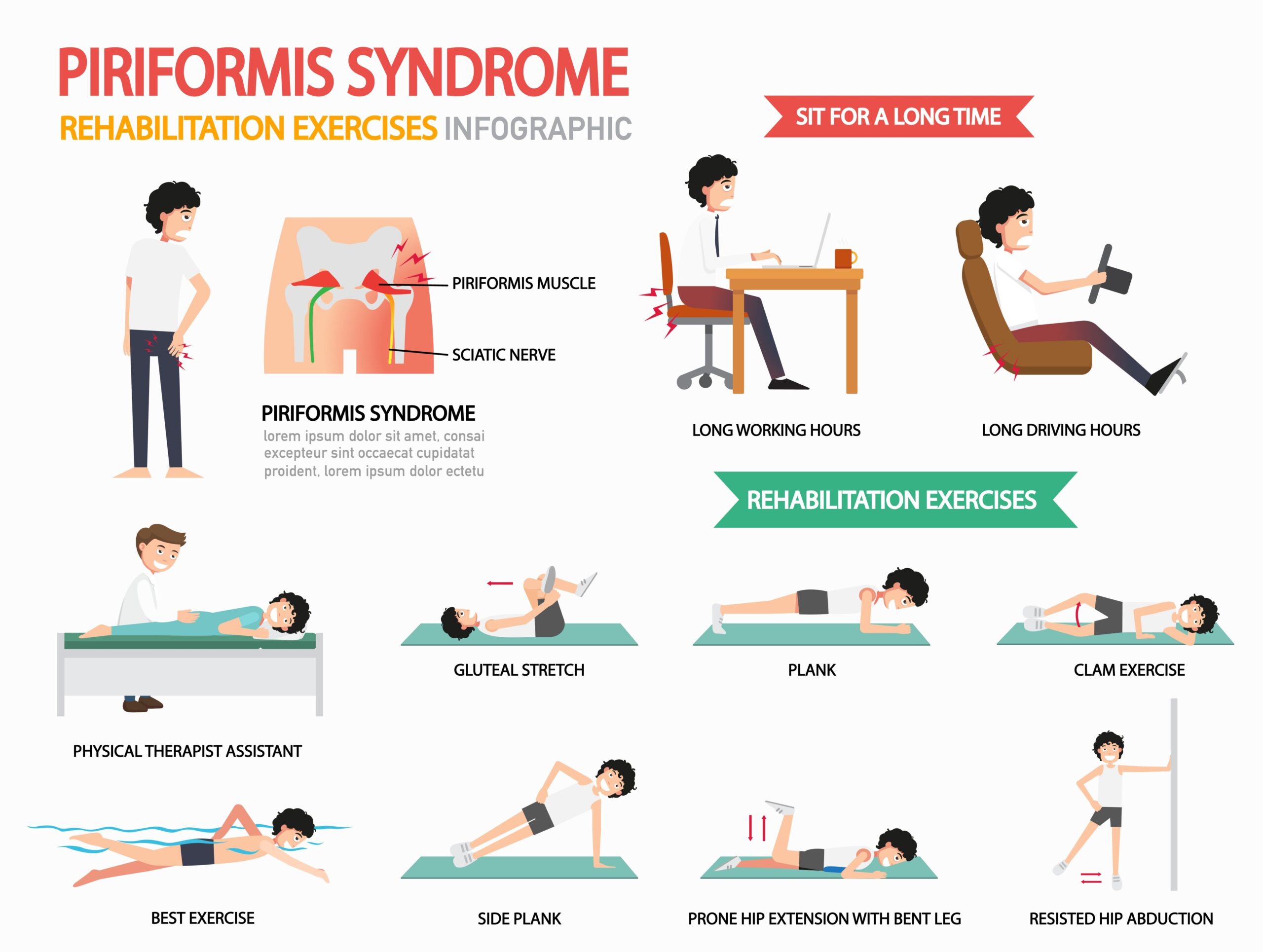
Exercise is the backbone of treating this syndrome. The goal is to stretch the muscle, strengthen the surrounding hip muscles, and reduce the pressure on the sciatic nerve. Fundamentally, we use these groups of movements:
- Piriformis stretch: While lying on your back, gently pulling the knee on the painful side toward the opposite shoulder. A mild sensation of tension is felt deep in the buttock, not sharp pain. (It is popularly known as the “figure-4 stretch.”)
- Hip stretching exercises: Movements that loosen the area around the hip, such as pulling both knees to the chest or laying the knees to the side.
- Gradual strengthening: The order here is important. We usually start with movements that work the hip under low load without straining the piriformis, such as the bridge and the clamshell; these target the weak hip muscles (gluteus medius) in particular. Once the muscles start working correctly, we gradually increase the load and move on to single-leg movements. The goal is to take the load off the piriformis and distribute it to the strong hip muscles.
- Self-massage with a foam roller: Placing the buttock on the roller and crossing the painful leg over the other, then slowly rolling back and forth, can reduce tension in the piriformis and surrounding tissues. Pausing briefly on tender spots is relieving—but without forcing into sharp pain.
- Nerve flossing: These are gentle movements aimed at helping the sciatic nerve glide more freely within the surrounding tissues. When done correctly, they help reduce the “adhesion” sensation of the nerve; but because they can increase irritation if done incorrectly/excessively, they must be learned under the supervision of a physiotherapist.
I generally recommend holding stretches for 20-30 seconds in each position and repeating them several times during the day. However, there is a very important rule here: exercises are done within the limit of pain, not by forcing through pain.
⚠️ If your pain increases during a stretch, do not do that movement.
A stretch should produce a mild sensation of tension deep in the buttock; it should not cause sharp, stabbing pain or pain radiating into the leg. If a movement increases your pain, it is wrong to force it thinking “it will loosen up if I do it”—because incorrect or excessive stretching can tighten the muscle further and increase spasm and nerve irritation. In such a situation, stop that movement and consult a physiotherapist about the correct technique.
Which of these movements is suitable for you, and the correct technique, should ideally be determined with the guidance of a physiotherapist. Because the same stretch that is relieving in one patient can—especially if applied incorrectly—increase the complaint in another.
⚠️ What you should avoid in piriformis syndrome
- Forcing stretches that cause sharp pain. “The more pain, the more benefit” is not true; excessive stretching increases spasm.
- Sitting motionless for long periods. The most common mistake is staying completely motionless because of the pain. On the contrary, short walking breaks every hour relax the muscle.
- Sitting on hard surfaces and with a wallet in the back pocket. Both put direct pressure on the nerve.
- Returning to sports without warming up and sudden twisting/jumping movements.
- Applying heavy exercises learned from the internet on your own, with incorrect technique, without seeing a physiotherapist.
Before starting exercises, the correct diagnosis must have been made; because a “piriformis exercise” applied to a lumbar disc herniation or another condition can do harm instead of good.
How Should You Lie Down With Piriformis Syndrome?
Nighttime pain is a complaint that piriformis patients frequently mention. Adjusting the sleeping position with a few small tweaks can provide marked relief:
- Do not lie on the painful side. Direct pressure compresses the muscle and the nerve.
- If you lie on your side, place a pillow between the two knees and slightly bend the upper leg; this reduces tension in the hip.
- If you lie on your back, place a thin pillow under the knees; the lower back and hip rest in a more neutral position.
- Avoiding sitting for long periods on hard surfaces and crossing your legs during the day also reduces nighttime pain.
If, despite these measures, there is pain that keeps you from sleeping and steadily increases, this may indicate that the condition is progressing; in such a situation, do not delay in seeking an evaluation.
Piriformis Syndrome in Women and During Pregnancy
Piriformis syndrome is more common in women than in men; the main reason is differences in the anatomy of the pelvis. Two periods in particular stand out:
- Pregnancy: With increased weight and hormonal changes, the pelvic ligaments loosen and the body’s center of gravity shifts. This new load distribution can strain the muscle and cause pain. Low back and hip pain is very common in pregnancy, and some of it originates from this muscle.
- The postpartum period: Changes in the pelvic muscles and one-sided carrying/sitting habits with the baby can trigger the condition.
The treatment approach in pregnancy is especially careful: the priority is appropriate stretching exercises, position modification, and physiotherapy; medication and interventional options are planned only with a doctor’s evaluation and in consideration of the pregnancy status. During this period, be sure to share your complaints with the doctor following your care.
When Is Surgery Needed for Piriformis Syndrome?
In piriformis syndrome, the first phase is always non-surgical treatment. However, if there is long-standing pain, a lack of response to physical therapy, or a serious decline in quality of life, surgery comes onto the agenda. In short, surgery is the last step, reserved for resistant cases.
I decide on surgery in the following situations:
- Severe pain lasting longer than 3 months
- Lack of response to non-surgical treatments including medication, exercise, and injection
- Support of nerve compression by EMG (nerve conduction study)
- Detection of marked hypertrophy or swelling in the muscle on MRI or US
The logic of the surgery is simple: the tendon portion of the muscle that presses on the nerve is released. There are two methods:
Open surgery
It is performed with an incision of about 7-8 cm at the back of the hip. In the great majority of patients, pain and numbness begin to decrease noticeably even right after surgery. One day after surgery, the patient can walk without support, sit, and shower; they usually return to daily activities within 10 days to 2 weeks.
Closed (endoscopic) surgery
It is much more comfortable than the open method. It is performed by entering through two holes of about 1 cm each; this means faster recovery and rehabilitation after surgery.
Surgical risks
Like any surgery, this too carries risks such as infection, bleeding, nerve injury, and anesthesia-related complications. However, when performed with the correct technique, piriformis surgery is an effective option and the rate of risk is low. Whether open or closed, the success rate is high.
For detailed information about the surgery and to determine the method suitable for you, you can reach us through our contact channels.
If the Piriformis Muscle Is Cut, Will There Be Loss of Function?
This is the most frequently asked question when surgery comes onto the agenda, and the answer is reassuring: no, there is no noticeable loss of function. The reason for this is the anatomy we described at the start of the article. The job of the piriformis is to rotate the hip and leg outward (external rotation), but there are 6 other muscles that perform this task. What’s more, the strongest external rotator of the hip is not the piriformis but the gluteus maximus—that is, the large, powerful muscle of the buttock. For this reason, when the piriformis is released, this task is taken over by the other muscles and the patient does not feel any loss in daily life.
How Is Piriformis Syndrome Prevented?
Piriformis syndrome is more common in desk workers, people who sit for long periods, and some athletes. With a few simple but effective habits, you can markedly reduce the risk:
- Don’t sit for long periods. Take short walking breaks every hour and correct your posture. Don’t sit with a wallet in your back pocket.
- Stretch regularly. Stretching movements targeting the piriformis provide a protective effect even at 5-10 minutes a day.
- Strengthen the hip and core muscles. Balance the load distribution with Pilates, yoga, or resistance exercises.
- Correct postural disorders. If needed, get posture training with the guidance of a physiotherapist.
- Don’t start sports without warming up. Warming up before sports and cooling down afterward protects the muscle.
- Avoid sudden, erratic movements. Sudden twisting, jumping, and heavy lifting can cause microtrauma in the muscle.
The Testimonial and Recovery Process of My Patient Who Had Piriformis Surgery
Listening to the experiences of patients who have had surgery is one of the best ways to manage expectations for patients wondering about the process. In the video below, you can watch, in her own words, the recovery process of a patient on whom I performed piriformis release surgery after a lengthy treatment process.
In general, the recovery process after surgery (especially with the closed/endoscopic method) is fast:
- Hospital stay: Usually 1 night; in some patients, same-day discharge.
- Walking: A few hours after surgery, the patient stands up, walks, and manages their own toilet needs.
- Return to work: Desk workers within 7-10 days, manual workers within 3-4 weeks.
Piriformis Syndrome vs. Lumbar Disc Herniation Differential Diagnosis Table
| Feature | Piriformis Syndrome | Lumbar Disc Herniation |
|---|---|---|
| Center of the pain | Deep in the buttock; markedly increases with sitting | Radiates from the lower back into the leg; increases with bending forward |
| Neurological findings | Usually not prominent | Band-like numbness, loss of strength, and possible foot drop in the region the nerve supplies |
| Triggering movement | Prolonged sitting, internal rotation of the leg | Bending forward, coughing, straining |
| Characteristic clue | Inability to sit for more than 15-20 minutes | Pain descending below the knee, to the foot |
| Imaging | MRI usually normal; diagnosis made by exclusion | Disc protrusion / nerve-root compression on MRI |
| Prominent treatment | US-guided piriformis injection, stretching | Treatment directed at the lower back; epidural in cases that require it |
Note: In daily practice, a lumbar disc herniation and the piriformis can also be present together. That is why this table is not a decision tool on its own, but a roadmap that must be evaluated together with the examination. For interventional options directed at a lumbar disc herniation, you can review our lumbar disc herniation injection treatment page.
Conclusion
Piriformis syndrome is a condition that occurs when the piriformis muscle deep in the buttock presses on the sciatic nerve, showing itself with sciatica-like pain and numbness. Its most insidious aspect is that it exactly mimics a lumbar disc herniation and usually does not show up on MRI—which is why many patients lose time at the wrong clinic, with the wrong diagnosis.
The good news is this: when the correct diagnosis is made, treatment is largely non-surgical. With activity modification, stretching exercises, and, when needed, US-guided injection treatments, most early cases improve markedly within 4-8 weeks. Surgery is the last step, which we reserve only for resistant cases, and it leaves no loss of function.
As an endurance athlete and orthopedic specialist, what I emphasize most is early and accurate diagnosis. If, despite a clean lumbar MRI, you have pain radiating from the hip down the leg that won’t let you sit, don’t ignore it. For an evaluation and a treatment plan suited to you, get in touch with us—let’s take the first step together toward a pain-free and active life.
Frequently Asked Questions About Piriformis Syndrome
How long does piriformis syndrome take to resolve?
In mild to moderate cases that are caught early and managed correctly, most patients see marked improvement within 4-8 weeks with stretching exercises, activity modification, and a US-guided injection when needed. In chronic cases lasting longer than 3 months, recovery takes longer. It is not right to give an exact number of days; seeking care early is the most important factor in shortening recovery.
My lumbar MRI was clear but my leg hurts—could it be piriformis?
Yes, this is the most typical scenario for piriformis syndrome. Piriformis is a diagnosis of exclusion and usually does not show up directly on MRI. If your lumbar MRI is clear and you still have sciatica-type pain radiating from the hip down the leg, a lumbar disc herniation has been ruled out and the remaining picture strongly points to the piriformis. The definitive distinction is made with an examination and, when needed, a US-guided diagnostic injection.
How is piriformis syndrome distinguished from a lumbar disc herniation?
The most valuable clue is the patient’s inability to sit for long periods: piriformis patients cannot sit for more than 15-20 minutes, especially on hard surfaces, and constantly change position. With a lumbar disc herniation, clear neurological findings such as marked loss of sensation, weakness in the toes, or foot drop, and pain descending below the knee are more prominent. Where the pain starts is not a reliable distinguishing factor on its own.
Do injections help in piriformis syndrome—cortisone, Botox, or PRP?
Injections are helpful when they are placed precisely into the muscle under ultrasound (US) guidance rather than blindly. Cortisone is the most commonly used option; it acts quickly but its effect is relatively short-lived, reducing pain and making it easier to start exercising. Botox has the strongest level of evidence among injections for piriformis syndrome; controlled studies show it can give superior results compared to cortisone plus local anesthetic, and its effect lasts longer (about 3-6 months), so it is preferred in resistant spasms. PRP is a method prepared from the person’s own blood that targets tissue repair; the evidence for it in the piriformis is still new and limited, so it is considered for selected resistant cases that have not responded to established treatments (exercise, cortisone, Botox). The right choice is determined individually based on the patient’s clinical picture.
If the muscle is cut during piriformis surgery, will there be loss of function?
No. The job of the piriformis muscle is to rotate the hip and leg outward (external rotation), and there are 6 other muscles that perform this task. Moreover, the strongest external rotator is the gluteus maximus—that is, the large muscle of the buttock. For this reason, when the piriformis is released, this task is taken over by the other muscles and patients do not experience any noticeable loss of function.
Does piriformis syndrome resolve on its own?
In mild cases, symptoms can ease within weeks with activity modification, ice application, appropriate medication, and regular stretching exercises, and depending on the picture they may regress. However, as long as strenuous movements and prolonged sitting continue, the pain returns. Chronic cases lasting longer than 3 months are not expected to resolve on their own.
Stretching increases my pain—should I still do it?
No. A stretch should produce only a mild sensation of tension deep in the buttock; it should not cause sharp, stabbing pain or pain radiating into the leg. If a movement increases your pain, do not do it and do not insist that “it will loosen up if I push through,” because incorrect or excessive stretching can tighten the muscle further and increase spasm and nerve irritation. Exercises are done within the limit of pain. The best approach is to stop the movement that causes pain and consult a physiotherapist about the correct technique.
How should I lie down with piriformis syndrome, and which position brings relief?
Generally, not lying on the painful side is the most relieving. Placing a pillow under the knees when lying on your back, or placing a pillow between the two knees and slightly bending the upper leg when lying on your side, reduces tension in the piriformis muscle. Avoiding sitting for long periods on hard surfaces and crossing your legs also lowers the pain.
Which doctor should I see for piriformis syndrome?
Piriformis syndrome is primarily evaluated by an orthopedics and traumatology specialist and a physical medicine and rehabilitation (PM&R) specialist. In situations where a lumbar disc herniation and other neurological causes need to be ruled out, neurology may also be involved in the process. An accurate diagnosis is the most critical step of treatment.
Does piriformis syndrome show up on MRI?
Most of the time it does not show up directly. In only about 10-15% of patients with a confirmed piriformis diagnosis is the muscle seen enlarged compared to the opposite side on pelvic MRI. The real value of MRI is to rule out causes that produce a similar picture, such as a lumbar disc herniation; that is, it shows what is not present, and the examination completes the rest of the picture.
Which examination tests are performed for piriformis syndrome?
Special tests are used that temporarily increase pressure on the nerve by stretching or contracting the piriformis muscle and reproduce the familiar pain. The most commonly used are the FAIR test (flexing the hip and knee and internally rotating the leg), the Freiberg test (internal rotation of the leg), the Pace test (resisted abduction of the legs), and the Beatty test (raising the upper leg while lying on the side). A single positive test is not sufficient; what really matters is that several tests together reproduce the pain. These tests are evaluated together with the patient’s history and other findings.
What is MR neurography, and is it needed in piriformis syndrome?
MR neurography is a special MRI technique tuned to image the nerve itself directly. While a standard MRI mostly shows bone and general soft tissue, MR neurography can reveal signs of thinning, thickening, or compression in the sciatic nerve. It is not needed for every patient and is not available at every center; it can be a valuable guide in resistant cases where the diagnosis is unclear and that do not respond to non-surgical treatment. Even so, even the most advanced imaging does not replace the examination—it complements it.
Can you walk with piriformis syndrome?
The type of walking is decisive. Short, gentle walks are beneficial and are generally recommended because they increase blood circulation and relax the muscle. Long, brisk walks, on the other hand, can tire the muscle and increase spasm, so they are not recommended during an active pain period. It is best to start walking slowly, increase the duration gradually, and stretch after walking. If your pain increases markedly with walking, shorten the duration and consult your doctor.
In piriformis syndrome, is heat or cold application better?
Both have their place, and it depends on timing. Because muscle spasm is usually prominent in piriformis syndrome, heat application (a hot water bottle, a warm shower) is more effective at relaxing the muscle; it is preferred especially in long-standing complaints. In newly started acute pain or after exercise, cold application (ice, 15-20 minutes, 2-3 times a day) helps reduce swelling and pain. A practical rule: heat before exercise, cold after exercise.
Is crossing your legs harmful in piriformis syndrome?
Yes, it is a habit to avoid during the active period. Crossing your legs stretches the hip muscles, is a position that contracts the piriformis muscle, and can trigger pain by increasing pressure on the sciatic nerve passing beneath it. Sitting for long periods on hard surfaces and sitting with a wallet in the back pocket similarly compress the nerve; avoiding these three habits noticeably reduces the pain.
Why is piriformis syndrome more common in women?
Piriformis syndrome is more common in women than in men; the main reason is differences in the anatomy of the pelvis. The wider structure of the female pelvis can strain the muscle by affecting load distribution across the hip and the way the piriformis muscle works. In addition, the increased weight, shifted center of gravity, and loosened pelvic ligaments during pregnancy can place extra load on the piriformis muscle and trigger the condition.
Are piriformis tightness and piriformis syndrome the same thing?
No, and the difference between them determines the treatment. With piriformis tightness, pain is felt only locally, deep in the buttock; there is no radiation into the leg, numbness, or tingling—meaning the nerve is not yet involved. In piriformis syndrome, the muscle presses on the sciatic nerve; pain radiates into the leg and nerve findings such as numbness and tingling are added. If tightness is not treated in time, it can progress to the syndrome. The ideal treatment window is the tightness phase, before the nerve pain radiating into the leg begins; in this phase, stretching exercises and, when needed, a series of US-guided injections (usually 3 doses one month apart) are applied.
Are deep gluteal space syndrome and piriformis syndrome the same thing?
Not exactly the same; piriformis syndrome is the most common subtype of deep gluteal space syndrome. Deep in the buttock, the sciatic nerve can be compressed not only by the piriformis muscle but also by other muscles, bands of connective tissue, or blood vessels. This entire picture is called deep gluteal space syndrome. That is why, in hip-leg pain that persists despite a clean lumbar MRI, an examination that evaluates the whole region without fixating on a single muscle is important.
Thank you for reading our article. If you wish, you can return to the utkuerdemozer.com homepage or get in touch with us for your questions.