Patellar dislocation, like any joint dislocation, is an orthopedic emergency. When the kneecap (patella) is dislocated, severe pain, deformity, and restricted movement occur. It must be reduced (relocated) as soon as possible. However, the actual treatment plan begins after this initial intervention.
Добавьте нас в предпочитаемые источники GoogleНаши новые статьи будут выше в вашем поискеДобавитьThe patient’s age, whether it is a first-time or recurrent dislocation, the presence of cartilage damage or loose bodies inside the joint, and structural abnormalities such as malalignment (e.g., tibial tubercle–trochlear groove distance over 20 mm) should be carefully assessed. The decision for surgery is shaped by these parameters; not every patellar dislocation requires surgery, and not every case is treated with the same surgical procedure.
What Is Patellar Dislocation?
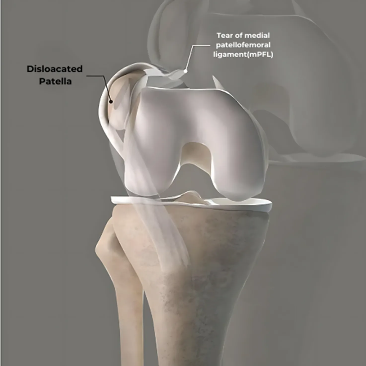
The patella is an important bone that allows the knee to bend and straighten. Normally, it moves only up and down and does not shift sideways. However, during uncontrolled movements such as sudden twisting, falling, or knee rotation, the patella can move outward from the trochlear groove of the femur. This is called patellar dislocation.
Patellar dislocations most often occur laterally (to the outside), known as lateral patellar dislocation. Following a dislocation, the MPFL (medial patellofemoral ligament) is usually injured. If it does not heal properly, the risk of recurrence increases. This can lead to ligament injuries, cartilage damage, and in the long term, anterior knee pain and patellar instability.
Why Does the Patella Dislocate?
Patellar dislocation most commonly occurs during sports activities, especially in non-contact twisting injuries of the knee. However, in contact sports such as football or basketball, a direct blow to the knee area can also cause patellar dislocation. Almost always, the dislocation occurs to the outside (lateral). Rarely, in congenital structural abnormalities, the patella may dislocate medially (to the inside), usually due to developmental issues during fetal growth.
The quadriceps tendon, which is the common tendon of the quadriceps muscles, attaches to the upper part of the patella, and the patellar ligament attaches to the lower part. These structures keep the patella stable within the femoral trochlea. The most critical soft tissue structure ensuring this stability is the medial patellofemoral ligament (MPFL), which prevents the patella from shifting outward. This ligament runs between the inner-upper edge of the patella and the medial femoral condyle.
During a patellar dislocation, the MPFL usually tears at its femoral attachment site. If this tear does not heal properly after the first dislocation, it poses a significant risk for recurrent patellar dislocations. In most patients with recurrent dislocations, MPFL insufficiency is the underlying cause.
In some cases, the patella may relocate into the trochlea spontaneously after dislocating. However, if it does not return to its position, it must be reduced by an orthopedic surgeon. Since dislocations can be accompanied by loose bodies, cartilage damage, or bone bruising, imaging should be performed in every case to assess the need for further treatment.
Diagnosis of Patellar Dislocation
Patellar dislocation is usually diagnosed easily through clinical examination. Patients often report sudden severe pain, restricted movement, and visible deformity of the knee. The patella typically shifts laterally and can be detected during careful examination. Sometimes, the patella may spontaneously relocate; however, ligament and cartilage damage risk remains.
Imaging methods are important in diagnosis:
- Direct X-ray: Evaluates patellar position, associated fractures, and anatomical predisposition (e.g., patella alta, trochlear dysplasia).
- Magnetic Resonance Imaging (MRI): Especially recommended after the first dislocation. It clearly shows MPFL tears, cartilage injury, bone bruising, and loose bodies.
- Computed Tomography (CT): Used in selected cases to evaluate bony anatomy contributing to patellar instability. TT–TG (tibial tubercle–trochlear groove) distance measurement, femoral and tibial rotation, and version analysis are performed via CT. These measurements are critical for predicting recurrence risk and guiding surgical planning.
The key in diagnosis is not only to confirm the dislocation but also to identify underlying structural risk factors and soft tissue damage, as these determine an individualized treatment plan.
Factors That Increase the Risk of Patellar Dislocation
Patellar dislocation can occur at any age, but certain anatomical and biomechanical factors increase susceptibility. If these are not evaluated after the first dislocation, the patella may repeatedly dislocate, causing permanent knee damage.
- History of Patellar Dislocation
Once the patella has dislocated, the risk of recurrence increases significantly. MPFL damage or incomplete healing predisposes to repeated dislocations. - Patella Alta
A patella positioned higher than normal enters the trochlear groove later during knee flexion, increasing the risk of lateral displacement. - Trochlear Dysplasia
The femoral trochlea is the groove where the patella sits. A shallow or flat groove facilitates lateral patellar shift. Trochlear dysplasia is a common structural abnormality in patients with patellar dislocation. - Bony Alignment and Axis Deformities
Malalignment between the hip, knee, and ankle (e.g., genu valgum – knock-knee, or genu varum – bowleg) alters forces on the patella and increases dislocation risk. Rotational deformities, such as increased femoral anteversion, should also be evaluated. CT imaging is used for rotational and TT–TG distance measurements in such cases. - Female Gender
As with anterior cruciate ligament injuries, patellar dislocations are more common in women, partly due to greater ligamentous laxity.
Symptoms of Patellar Dislocation
Patellar dislocation is a painful condition that causes loss of function. It typically occurs after acute trauma and presents with distinct clinical signs:
- Severe Pain: The patella moves laterally out of the trochlea, damaging surrounding soft tissues, which almost always causes intense pain until it is reduced.
- Swelling: Damage to intra-articular tissues and small blood vessels can cause bleeding into the joint (hemarthrosis), leading to visible swelling.
- Deformity: When the patella is displaced laterally, an obvious asymmetry is seen at the front of the knee. The patella is not in its normal position, which is both visible and functionally limiting. Patients often hold the knee in a flexed position for comfort.
- Restricted Movement and Weakness: Extension becomes painful, and quadriceps strength decreases, leading to a feeling of weakness in the leg.
- J Sign: A key finding in physical examination for patellar instability. As the patient moves from knee extension to flexion, the patella suddenly shifts outward in a pattern resembling the letter “J.” This is significant in recurrent dislocations and indicates underlying malalignment.
How Is Patellar Dislocation Treated?
Patellar dislocation is an orthopedic emergency that requires immediate intervention. After a dislocation, the patient experiences severe pain, swelling, deformity, and restricted movement in the knee. These symptoms will not significantly improve until the patella is relocated.
The treatment process has two phases: initial emergency management and long-term treatment planning based on the extent of injury.
1. Acute Management (Initial Intervention)
Once a patellar dislocation is diagnosed, X-ray imaging is performed to evaluate the position of the patella and check for any associated fractures. If the patella is still dislocated, it is carefully relocated (reduction) by an orthopedic surgeon. Afterwards, MRI is used to assess intra-articular structures (cartilage, ligaments, loose bodies) in detail.
2. Non-Surgical Treatment Options
If the first dislocation is not accompanied by significant cartilage damage or loose bodies inside the joint, the following protocol is applied:
- Ice application: Applied for 15–20 minutes several times a day to reduce swelling and pain.
- Painkillers: Particularly effective in controlling pain and inflammation in the first days.
- Knee brace: Used for a certain period to stabilize the patella; duration is determined according to the clinical condition.
- Physiotherapy: Starts with controlled movements. The main goal is to strengthen the quadriceps, especially with closed-chain exercises. Hip and core strengthening exercises are also included in the program.
- Joint fluid drainage (arthrocentesis): If there is significant fluid accumulation, draining it reduces pain and facilitates the transition to physiotherapy.
3. When Surgery Is Required
Surgical treatment should be considered in the following situations:
- Severe MPFL tear or avulsion from bone
- Deep cartilage defects
- Presence of loose bodies inside the joint
- Anatomical abnormalities such as trochlear dysplasia or patella alta that cause recurrent dislocations
- Second or subsequent dislocations
The goal of surgery is to repair or reconstruct the structures that stabilize the patella. One of the most common procedures is medial patellofemoral ligament (MPFL) reconstruction, which is often performed together with tibial tubercle osteotomy.
Patellar Dislocation Surgeries
In some cases, surgery is absolutely necessary for patellar dislocation. If there is a loose cartilage fragment (loose body) inside the joint or cartilage damage requiring intervention, surgery is indicated regardless of the number of dislocations or the patient’s age. In such cases, both patellar stabilization surgery and cartilage surgery are planned simultaneously.
If there is no cartilage problem but imaging (CT and MRI) reveals anatomical factors that increase recurrence risk, surgery is again considered. The most commonly performed procedures in these patients include:
Cartilage Surgeries
If a loose cartilage fragment (loose body) forms inside the joint during dislocation, surgical treatment is mandatory.
During arthroscopic knee surgery:
- If the fragment is small, it is removed.
- If the fragment is large, it is fixed back to its original site with screws.
The aim of these surgeries is to preserve the knee’s cartilage surface and prevent future osteoarthritis.
MPFL Reconstruction Surgery
The medial patellofemoral ligament (MPFL), which often tears during dislocation, is one of the key stabilizing ligaments preventing the patella from shifting outward.
In patients with MPFL tears, this ligament needs to be reconstructed. This is usually performed in the same session after diagnostic arthroscopy.
(You can click here to visit our MPFL reconstruction surgery page.)
Tibial Tubercle Osteotomy (Fulkerson Osteotomy)
CT imaging and TT–TG distance measurement determine whether the patella is anatomically predisposed to lateral dislocation.
If the TT–TG distance is over 20 mm, tibial tubercle osteotomy may be required to realign the patella. In this procedure, the tibial tubercle bone is surgically repositioned.
(You can click here to visit our tibial tubercle osteotomy surgery page.)
Surgery Videos
You can watch surgeries performed by Op. Dr. Utku Erdem Özer from the following links:
- Click here to watch my MPFL reconstruction surgery video
- Click here to watch my tibial tubercle osteotomy surgery video
Recommended Exercises After Patellar Dislocation
The exercise program after patellar dislocation aims to strengthen the muscles around the knee and improve joint stability. Strengthening the quadriceps and hamstrings in particular helps prevent the patella from dislocating again.
Goals of the Exercises
- Improve coordination of the muscles around the knee
- Maintain knee range of motion
- Reduce load on the patellofemoral joint
- Increase knee stability
- Minimize the risk of recurrent dislocation
Recommended Exercises
- Straight Leg Raise
Lie on your back with one leg straight and the other knee bent. Slowly lift the straight leg to about 45 degrees, then lower it.
Purpose: Strengthen the quadriceps muscle. - Quad Set (Isometric Quadriceps Exercise)
Place a rolled towel under your knee. Tighten your quadriceps, pressing the knee down against the towel. Hold for 5 seconds, then release.
Purpose: Safely activate the quadriceps muscle in the early postoperative period. - Heel Slide
While lying on your back, slowly slide your heel toward your hips, then return to the starting position.
Purpose: Maintain knee range of motion. - Wall Slide
Stand with your back against a wall, feet shoulder-width apart. Slowly slide down until your knees are bent 30–45 degrees, then return to the starting position.
Purpose: Activate both the quadriceps and gluteal muscles. - Step-Up
Step up and down on a low platform using one leg at a time.
Purpose: Strengthen the quadriceps, hip muscles, and stabilizing muscles together.
Note: These exercises should be performed under the supervision of a physiotherapist and tailored to the patient’s current condition. Incorrectly performed exercises may cause the patella to dislocate again.
Frequently Asked Questions About Patellar Dislocation
What is recurrent patellar dislocation?
If the patella dislocates more than once, this is called recurrent patellar dislocation. With each dislocation, the joint cartilage sustains more damage, and knee stability progressively worsens. For this reason, surgery is the definitive treatment for recurrent patellar dislocation. Before surgery, a detailed evaluation should be performed to identify all anatomical causes of instability, and the surgical plan should aim to address all of them in a single procedure.
How long does it take to recover from a patellar dislocation?
Recovery time depends on the severity of the dislocation and accompanying injuries. First-time dislocation without structural damage usually recovers within 2–4 weeks. Recurrent dislocations or cases with cartilage/ligament injury may take 6–8 weeks or longer. If surgery is required, full recovery can take 3–6 months.
What happens if patellar dislocation is not treated properly?
If a patellar dislocation or subluxation is not treated appropriately in the early period, it can lead to permanent knee damage. Recurrent dislocations predispose the joint to cartilage damage and patellofemoral arthritis. Long-term consequences include recurrent dislocations (40–60% risk without treatment), cartilage damage (lesions in over 60% of recurrent cases), patellofemoral arthritis (up to 50% risk within 10 years), quadriceps weakness (up to 30% reduction in vastus medialis activity), restricted movement, and need for more complex surgery.
How is a dislocated kneecap treated?
Treatment depends on the patient’s age, recurrence status, and joint damage. First intervention involves transporting the patient safely, reduction by an orthopedic specialist, and imaging (MRI/CT). Non-surgical treatment includes bracing, ice, physiotherapy, and strengthening exercises, especially after first-time dislocation. Surgery is necessary if there is a loose body, recurrent dislocation, or anatomical abnormalities. Surgical options include MPFL reconstruction, Fulkerson osteotomy, and cartilage repair. Strengthening quadriceps, hamstrings, and hip abductors is crucial in prevention and recovery.
What happens if a dislocated kneecap is left untreated?
Untreated patellar dislocations can lead to recurrent dislocations, cartilage damage, patellofemoral arthritis, muscle weakness, restricted function, and more complex surgeries in the future.
Does treating a dislocated kneecap always require surgery?
Not always. Surgery is mandatory if there is a loose body or cartilage injury. If imaging shows risk factors such as trochlear dysplasia or MPFL laxity, surgery like MPFL reconstruction or Fulkerson osteotomy may be performed. If no risk factors are present, rehabilitation may be sufficient. After a second dislocation, surgery is usually inevitable.
Why does the kneecap dislocate?
The kneecap can dislocate due to sudden twisting, trauma, or weak muscles. Structural abnormalities such as trochlear dysplasia, patella alta, or increased Q-angle also increase risk.
Can a dislocated kneecap heal on its own?
No. A dislocated kneecap typically requires closed reduction by an orthopedic specialist. Associated cartilage or ligament injuries should also be evaluated.
Why does the kneecap usually dislocate outwards?
Anatomically, the patella is more prone to lateral dislocation due to groove shape and biomechanical factors. Weak muscles, patella alta, increased Q-angle, or excessive TT–TG distance increase lateral dislocation risk.
What happens if a dislocated kneecap is not surgically repaired?
Without surgical repair, patients face recurrent instability, cartilage damage, and early-onset patellofemoral arthritis, leading to reduced quality of life.
Is MRI necessary after a kneecap dislocation?
Yes. MRI helps detect cartilage, ligament, and soft tissue injuries, especially MPFL tears or cartilage fractures after the first dislocation.
When is surgery recommended after a kneecap dislocation?
Surgery is recommended after a second dislocation or if the first dislocation is accompanied by cartilage fracture (loose body). It may also be required if TT–TG distance exceeds 20 mm or if MPFL tears are present.
What surgeries are performed for kneecap dislocation?
The main surgeries are MPFL reconstruction, Fulkerson osteotomy, and arthroscopic cartilage repair. The choice depends on imaging and recurrence history.
How long does it take to recover from a kneecap dislocation?
Non-surgical cases usually recover within 6–8 weeks. Surgical recovery takes 3–6 months, depending on the procedure, with physiotherapy included.
Which exercises should be done after a kneecap dislocation?
Exercises should strengthen the quadriceps (especially vastus medialis), hamstrings, hip abductors, and core. Closed-chain exercises are recommended under physiotherapist supervision.
How can a kneecap dislocation be prevented?
Muscle balance correction, strengthening exercises, maintaining flexibility, and using knee braces when necessary can reduce risk. If structural abnormalities are present, surgery may be needed to correct them.
Conclusion
Patellar dislocation is a serious joint injury that can cause pain and functional loss. During dislocation, the ligaments and muscles of the joint may be strained or torn, leading to severe pain, swelling, and restricted movement. Therefore, when the patella dislocates, an orthopedic specialist should be consulted immediately.
After initial intervention, imaging methods such as X-ray and MRI are used to evaluate intra-articular damage. The treatment plan is shaped according to the severity of the injury, accompanying structural problems, and the patient’s overall health. Conservative methods such as ice application, pain medication, knee brace use, and physiotherapy may be sufficient in mild cases, while recurrent dislocations or cartilage damage may require surgical treatment.
Recovery time after surgery is individual. In mild cases, a return to normal life may be possible in 2–4 weeks, while in more severe injuries, recovery may take over 6–8 weeks. During this period, following medical advice, adhering to the exercise program, and being patient are critical.If you have questions about patellar dislocation or want more details about treatment options, you can contact us via WhatsApp or through our website at utkuerdemozer.com.