Knee osteonecrosis is a serious condition that occurs when blood flow to the bone tissue in the knee joint decreases, which, if left untreated, can lead to joint damage. In everyday language, this condition is also known as bone decay due to insufficient blood supply to the bone tissue.
Add us as a preferred source on GoogleSee our new articles higher in your searchesAddLong-term corticosteroid use, excessive alcohol consumption, autoimmune diseases, sickle cell anemia, and conditions that increase blood clotting are among the most important risk factors.
In addition, most cases are associated with meniscus root tears and subchondral insufficiency fractures of the articular cartilage. Magnetic Resonance Imaging (MRI) is usually required for early diagnosis.
Treatment generally begins with addressing the underlying cause and reducing the load on the joint. If a meniscus root tear is present, surgical repair should be performed first. In recent years, especially in the early stages, intraosseous bioplasty and bone marrow-derived stem cell applications have shown successful results.
What Is Knee Osteonecrosis?
Knee osteonecrosis most often develops in the inner portion of the femur (medial femoral condyle) and is characterized by impaired blood supply and insufficiency fractures.
In the early stage, bone edema and microfractures occur. As the process progresses, the bone weakens, can no longer support the cartilage surface, and collapse of the joint may occur.
If left untreated, permanent knee osteoarthritis and loss of mobility can develop.
Causes of Knee Osteonecrosis
In the past, many cases of spontaneous osteonecrosis with an unknown cause were later understood to be due to undetected meniscus root tears.
These tears can occur during daily activities such as squatting, cleaning, gardening, or during sports like skiing. The meniscus shifts outward, losing its protective function, and causes excessive loading on the joint.
You can read my article on meniscus root tears here.
Other factors that may play a role in the development of knee osteonecrosis include:
- After meniscus surgery
- Long-term corticosteroid use
- Inadequate blood supply to bones
- Osteoporosis and advanced age
- Subchondral insufficiency fractures
- Meniscus root tears
- Excessive alcohol consumption (alcoholism)
- Decompression sickness (diver’s disease)
- Bone marrow replacement diseases (e.g., Gaucher disease)
- Sickle cell anemia
- Conditions that increase blood clotting (hypercoagulability)
- Systemic lupus erythematosus (SLE)
- Inflammatory bowel diseases
- Organ transplant patients
- Certain viral infections (CMV, hepatitis, HIV, rubella, measles, chickenpox)
- Protease inhibitors (a drug group used in HIV treatment)
- Trauma
Symptoms of Knee Osteonecrosis
Osteonecrosis typically develops in stages. The first symptom is often sudden and sharp pain felt on the inner side of the knee. This pain may begin suddenly after a specific activity, squatting, sports requiring deep knee flexion, or a minor injury.
In cases related to meniscus root tears, pain may initially be felt at the back of the knee, sometimes accompanied by a popping sensation. Within a few weeks, this pain shifts to the inner side of the knee, becoming more pronounced and intensifying due to direct contact between the joint cartilage and bone surface.
As the disease progresses, bearing weight on the affected knee and standing become more difficult, and moving the joint becomes painful.
Other symptoms include:
- Swelling in the front and inner side of the knee
- Tenderness around the knee
- Reduced range of motion
The disease may progress over several months to more than a year. Early diagnosis is extremely important, as some studies show that treatment initiated in the early stages can slow the progression and improve outcomes.
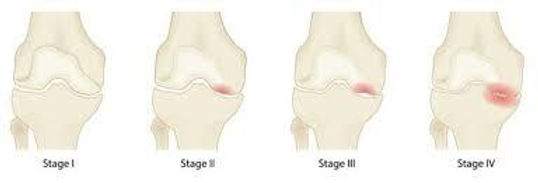
Stages of Osteonecrosis and Diagnostic Methods
- Stage 1: Initial stage, detectable only by MRI.
- Stage 2: Pre-collapse phase of the bone, detectable by X-ray and MRI.
- Stage 3: Partial collapse of the bone and formation of joint deformity.
- Stage 4: Advanced joint damage and deformity.
How Is Knee Osteonecrosis Diagnosed?
The diagnostic process begins with a detailed evaluation of the patient’s general health, medical history, and symptoms. A physical examination follows, during which swelling, warmth, redness, tenderness, joint range of motion, and stability are assessed. Pain upon weight-bearing and possible damage to surrounding muscles, tendons, and ligaments are also evaluated.
To confirm the diagnosis and determine the stage of the disease, imaging methods are used:
- X-ray: Shows changes in bone structure in advanced stages; usually normal in early stages.
- Magnetic Resonance Imaging (MRI): The most sensitive method in the early stage. Shows the extent of bone involvement and can detect osteonecrosis in the opposite knee before symptoms appear.
- Bone Scintigraphy: Used when necessary; a low-dose radioactive substance is administered intravenously, and areas of increased bone activity are identified—osteonecrosis typically shows increased uptake.
Treatment of Knee Osteonecrosis
Non-Surgical Treatment Methods
In the early stages, and when the affected bone area is small, non-surgical methods are preferred first. These include:
- Rest and avoiding weight-bearing on the affected knee; using crutches or a cane for a period if necessary
- Nonsteroidal anti-inflammatory drugs (NSAIDs), prescribed by a doctor, to reduce pain and inflammation
- In some cases, bisphosphonate therapy to slow bone loss
- Physical therapy, especially exercises to strengthen the thigh muscles and maintain joint range of motion; aquatic exercises if needed
- Regenerative treatments such as PRP and stem cell therapy
- Vitamin D and calcium supplementation
- Avoiding activities that place excessive load on the knee during daily activities
Surgical Treatment Methods
If pain persists despite non-surgical treatment or if there is a high risk of bone collapse, surgical options are considered:
- Arthroscopic debridement and microfracture technique
- Core decompression (reducing intraosseous pressure)
- Core decompression combined with osteochondral grafting or intraosseous bioplasty plus bone marrow-derived stem cell application
- Osteotomy (cutting the tibia or femur to change load distribution)
- In advanced stages, total knee replacement or partial knee replacement in suitable cases
Recommendations for Osteonecrosis Patients
- Avoid excessive load on the knee during daily activities
- Engage in regular, low-intensity walks
- Maintain proper weight control
- Follow a balanced, calcium-rich diet
Frequently Asked Questions (FAQ) About Osteonecrosis
Does knee osteonecrosis heal on its own?
In the early stages, it can sometimes heal spontaneously, but in most cases, treatment is needed to stop progression and prevent joint damage.
Is osteonecrosis progressive?
Yes, if left untreated, it can progress, leading to severe joint problems and cartilage loss.
Can knee osteonecrosis turn into cancer?
No, osteonecrosis does not turn into cancer.
Is surgery mandatory for osteonecrosis?
Surgery is not required in the early stages, but in advanced stages with collapse or severe cartilage damage, it may be unavoidable.
Is PRP effective in treating osteonecrosis?
PRP can help reduce symptoms and support healing in early-stage osteonecrosis.
What is intraosseous bioplasty and how is it used in osteonecrosis?
Intraosseous bioplasty is a procedure in which a special channel is created into the bone using the core decompression principle, and the space is filled with bone marrow-derived stem cells and biocompatible filler materials. The aim is to increase blood flow and strengthen the weakened bone.
What is the difference between intraosseous bioplasty and core decompression?
While core decompression only aims to reduce pressure and improve blood flow, intraosseous bioplasty also provides additional biological strengthening of the bone tissue.
How long is the recovery period after intraosseous bioplasty?
In most patients, gradual weight-bearing begins within 4–6 weeks. Full recovery time varies between 3–6 months depending on the extent of bone damage.
Can intraosseous bioplasty be combined with osteochondral grafting?
Yes, especially in cases with cartilage loss, it can be combined with osteochondral grafting to support the repair of the joint surface.