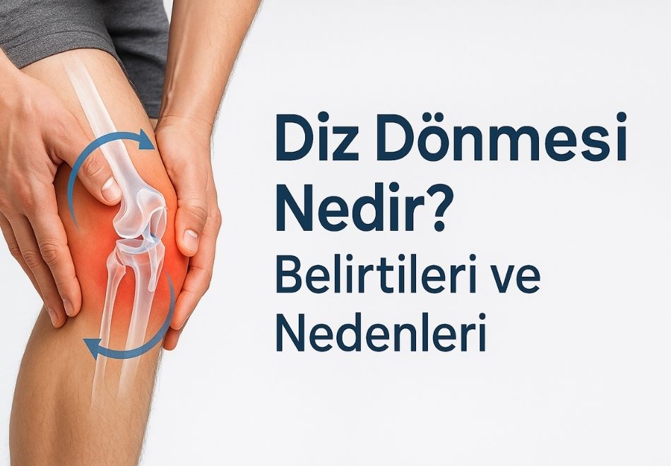
A knee sprain occurs when the knee joint suddenly twists around its axis, often leading to injuries in the anterior cruciate ligament, meniscus, or cartilage structures. It may happen during sports activities, while walking down stairs, or due to an unstable step.
Add us as a preferred source on GoogleSee our new articles higher in your searchesAddThe most common mechanism of a knee sprain is when the foot is fixed and the body rotates suddenly. It is especially common in athletes. Typical scenarios include sudden changes in direction while running, abrupt stopping, or landing from a jump when the foot makes contact with the ground (landing phase), causing rotational stress on the knee.
Which Structures Can Be Damaged After a Knee Sprain?
After a knee sprain, the most commonly injured structures are the soft tissues inside the knee. These structures are responsible for stability and smooth joint movement. During sudden twisting, these tissues can overstretch, tear, or become damaged.
1. Anterior Cruciate Ligament (ACL)
The ACL is the structure most often injured in knee sprains. It prevents forward sliding and rotation of the tibia. When the foot is fixed and the body rotates, the ACL can tear. A “popping” sound may be felt inside the knee at the moment of injury, followed by swelling within a short time.
2. Meniscus
There are two menisci in the knee — medial (inner) and lateral (outer). The lateral meniscus is the most affected in a twisting injury. Sudden external rotation of the knee can trap and tear the lateral meniscus. Meniscus tears often cause locking, sudden giving way, limited motion, and pain, especially during squatting.
3. Medial Collateral Ligament (MCL)
The MCL, located on the inner side of the knee, prevents inward bending. Force pushing the knee inward can partially or completely tear this ligament. It often accompanies ACL injuries.
4. Articular Cartilage
A twisting injury may damage the cartilage covering the joint surfaces. This is more likely if the knee hits something or if the bone surfaces inside the joint suddenly collide. Cartilage damage can predispose the knee to osteoarthritis later.
5. Bone Bruise (Bone Marrow Edema)
MRI after a knee sprain often shows bone marrow edema, indicating superficial bone trauma, which can cause prolonged knee pain.
In summary:
A knee sprain may look like a simple twist but can hide serious ligament, meniscus, or cartilage injuries. Early diagnosis and proper treatment can prevent long-term problems.
Symptoms of a Knee Sprain
The symptoms after a knee sprain vary depending on the severity of the injury and the structures affected. In mild sprains, there may be only short-term pain, while injuries to the ligaments or meniscus cause more pronounced symptoms.
Typical signs include:
- Sudden Knee Pain – Sharp, immediate pain right after twisting, usually localized to the inner or outer side of the knee.
- Popping or “Click” Sound – Common in ACL tears, caused by the ligament snapping.
- Swelling (Edema) – Noticeable swelling within hours, often due to bleeding (hemarthrosis) or fluid accumulation.
- Inability to Bear Weight – Difficulty putting weight on the affected leg, sometimes with a feeling of giving way.
- Limited Range of Motion – The knee may not fully bend or straighten; meniscus tears can cause locking or catching.
- Instability or Giving Way – Especially in ligament injuries, the knee may feel as if it will “slip out” or lose control.
In summary:
Pain, swelling, popping sounds, limited motion, and instability after a knee sprain may indicate a serious injury that requires prompt orthopedic evaluation.
How to Prevent a Knee Sprain
Although a knee sprain cannot be completely prevented, exercises that improve muscle strength, balance, and flexibility can significantly reduce the risk. Proper training programs strengthen the structures that protect the knee joint.
- Muscle Strength and Balance – Strong quadriceps and hamstrings are essential. Imbalance increases injury risk; both front and back thigh muscles should be equally strengthened.
- Hip and Core Strength – Weak hip and core muscles increase the load on the knee. Strengthening these muscles improves body mechanics, especially during sudden direction changes or jumps.
- Balance and Flexibility – Proprioceptive training (e.g., balance boards, single-leg exercises) improves joint stability. Adequate flexibility in hamstrings and calves prevents overstretching injuries.
- Knee Braces Do Not Prevent Sprains – Contrary to popular belief, braces only provide warmth and psychological support; they do not stop rotational injuries. The best protection is strong muscles and good coordination.
How Is a Knee Sprain Diagnosed?
A detailed examination and imaging are needed to identify injuries after a knee sprain.
- Physical Examination – Includes ligament stability tests (Lachman, McMurray, Thessaly, varus/valgus stress).
- MRI – The gold standard for detecting:
- ACL tears
- Meniscus tears
- Joint effusion
- Cartilage damage
- Bone bruises
In summary:
Accurate diagnosis is critical for effective treatment and recovery. A knee sprain should not be dismissed as a minor injury without orthopedic evaluation.
Treatment of a Knee Sprain
Treatment depends on which structures are injured and the severity.
Non-Surgical Treatment (for mild sprains, partial tears, minor cartilage damage):
- Rest
- Ice application (15–20 min several times daily)
- Compression and bracing
- Elevation
- Anti-inflammatory medication
- Physiotherapy and rehabilitation
Surgical Treatment (for severe injuries, especially in young and active individuals):
- ACL Reconstruction – Using grafts to replace a torn ligament
- Meniscus Repair or Removal – Depending on tear location and type
- Cartilage Repair Techniques – Microfracture, mosaicplasty, stem cell-supported repair
Which treatment is right?
It depends on the patient’s age, activity level, injury type, and expectations. Active young athletes often benefit from surgical repair, while older or less active patients may recover with non-surgical methods.